
Surgery


Are you looking for a reliable partner in your OR?
Then start to improve patient outcome with our crystal-clear visualisation and precise guidance before, during and after interventional procedures. Our ultrasound solutions integrate seamlessly into your surgical workflow, presenting an efficient, cost-effective, real-time and high-resolution imaging tool – with the necessary flexibility to adapt to your strategy.
- Get a clear picture with our superior ultrasound image quality.
- Enrich your interventions with advanced features like fusion and contrast imaging.
- Sense direct feedback with finger-grip probes during open HPB surgeries.
- Perform minimally invasive procedures with high resolution laparoscopic ultrasound.
- Find maximal flexibility with our robotic drop-in probes.
- Assess Colorectal findings prior, during and after surgery treatment
- Control your patient’s status with ultrasound-guided regional anaesthesiology.
Precise intraoperative ultrasound guidance for critical decisions – fast.
Ultrasound technologies for your clinical benefit
Combine highest ultrasound image quality with dedicated intraoperative probes and imaging features. Obtain quick and reliable information for all the steps of the procedure – from evaluating the condition, planning the strategy, guidance during the intervention up to validation of the outcome.
See every detail in high resolution
The combination of our image processing, probe technology and superior image optimisations permit to capture the subtlest of signals. Use Carving Imaging to obtain crystal-clear, sharp B-mode images for real-time surgical guidance, and pre- and post-operative anatomical assessment. A special feature is our Trapezoid, a virtual convex mode that enlarges your field of view with highest resolution.
Understand vascularisation of the operative site for safer outcomes
Now you can observe blood flow in even minute vessels with our highly sensitive Doppler and Colour modes like eFLOW or DFI. Assess vascular anatomy, find communicating veins, determine the adequacy of blood supply or detect a thrombus in your patient. This will guide your surgical procedure and improve post-operative follow-up.
Get a close look at micro-vasculature to localise, classify and treat lesions
Our contrast-enhanced ultrasound delivers vascular information beyond Doppler possibilities, with wideband pulse inversion and tissue reduction techniques. Differentiate between benign or malignant lesions, and stage tumours with confidence. To improve your surgical strategy, you can visualise isoechoic colorectal liver metastases and classify hepatocellular carcinomas with great sensitivity.
Take a harder look at soft tissue with elastography
Use strain elastography as digital palpation to display differences in tissue elasticity during the procedure – even in areas you can’t reach manually. Furthermore, you can easily quantify liver fibrosis with our point and 2D shear wave methods via the ARFI technique. All our intraoperative transducers incorporate an elastography function, so you can stay flexible and gather information from complimentary methods.
IOUS is an indispensable tool for all surgeons who want to perform hepatobiliary-pancreatic surgery at the highest level. Our intraoperative probes deliver the clearest images of even difficult-to-reach areas with a direct, tactile feedback. Combine this with advanced imaging modes to make informed operative decisions – for best patient outcomes.
Sense resection guidance in real time with our finger-grip probes
Originally designed for open surgery with renowned liver surgeon Prof. Masatoshi Makuuchi, our micro-convex probes can be easily griped between two fingers to allow palpation while scanning. The small bodies give you a direct sense for the tissue, and easily fit into a small operating field. Equipped with advanced imaging modes, they help you to guide resections in real time, and detect lesions during hepatic and pancreatic surgery.
Interpret the whole scan plane with linear T-shaped probes
For a wider field of view over large areas, make use of our T-shaped linear probes. Specifically designed for open surgeries, you can clearly visualise and follow anatomical structures and blood flows, easily interpret the scan plane and facilitate ablations. All the while Trapezoid, a virtual convex mode, further enlarges your field of view while maintaining the high resolution of the linear array.
Improve your procedures with advanced imaging modes
Our IOUS probes incorporate cutting-edge imaging modalities to help you succeed with better surgical outcomes: highly sensitive Colour and Doppler modes to visualize blood flow; contrast-enhanced ultrasound to stage tumours; elastography to differentiate tissue elasticity; or fusion imaging guidance with MRI and CT. Understand your patient’s status from various angles and make sure to know all you need to know during your procedure.
Step up to the next level of intraoperative navigation and guide your exact way through the body with our fusion imaging RVS. Combining CT, MRI or PET with real-time IOUS will help you locate lesions and target your liver resections with great precision. Adjust your preoperative strategy directly in the OR – for safe and accurate resection tailored to your patient.
Experience precise navigation with multimodality fusion imaging
Synchronize a reference image (from previous CT, MR, PET, or US scans) with live IOUS from our finger-grip probe with just one click. This enables you to guide your resection with great accuracy directly in the OR, and locate difficult-to-see structures – for example disappearing lesions after chemotherapy. Even complex liver resections or ablation treatments become easier, safer and more accurate.
Add even more insights with advanced ultrasound imaging modes
Combine fusion with other imaging modes in your real-time ultrasound scan. Switch on Colour Flows and Elastography, or enhance microvasculature with contrast agents to obtain a complete understanding of your patient’s condition. This truly multiparametric intraoperative solution will help you to be more efficient, with accurate positioning of RF ablation needles. Stay flexible, react quickly and make informed decisions.
Bring your pre-operative planning of liver resections directly in the OR
Integrate your pre-operative planning, including all anatomical information and volumetry done with Synapse 3D Liver Analysis*, into our fusion imaging solution. With this unique combination, you can reference clearly and in real time all resection lines, lesions and margins for a safe and accurate guidance through this complex procedure.
*Liver Analysis module from Synapse 3D is a product of Fujifilm Medical Co., Ltd in Japan.
Our flexible and easy-to-use laparoscopic ultrasound probes support efficient keyhole surgeries in HPB, urology, coloproctology, gynecology and more. And here, less is more: less patient trauma, less complications, less scarring and less recovery time – for better patient outcome.
Combine optimal organ contact with high-resolution imaging
See beyond the surface into the organ anatomy with our laparoscopic probe. Its linear array allows firm contact with the organ, so you can overcome the lack of tactile sensation and capture immediate, real-time feedback throughout the procedure. At the same time, high-resolution imaging clearly differentiates tissue structures to locate lesions, and very sensitive Colour and Doppler modes help you to obtain detailed vascular information.
Image organs from all angles with flexible and slim design
Articulate the tip of the probe in four different directions. This flexibility – combined with a slim body that easily fits through a trocar – allows you to investigate the operation site from any angle. Localize tumours, grasp the positional relationship with surrounding vessels or search for residual lesions. All the while Trapezoid, a virtual convex mode, enlarges your field of view while maintaining the high resolution of the linear array.
Improve your laparoscopy with advanced imaging modes
Our laparoscopic probe incorporates cutting-edge imaging modalities to help you succeed with better surgical outcomes: apply contrast-enhanced ultrasound to stage tumours; and use elastography to differentiate tissue elasticity and improve lesion characterization. You can understand your patient’s status from various angles and make sure to know all you need to know during your procedure.
The advantages of robotic-assisted surgeries are evident: steady, small, flexible, objective and accurate, improving the outcome of delicate procedures. Seamlessly integrate our drop-in ultrasound probes to identify key landmarks and guide successful treatment – with direct control, great precision and dexterity.
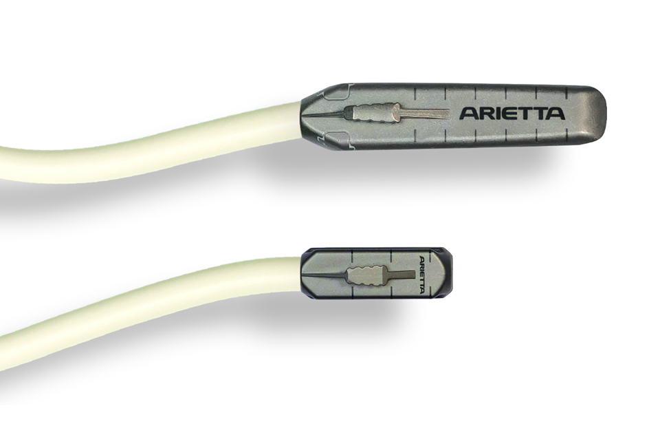
Get a 360° surgical flex for optimal in-situ usability
Our intraoperative drop-in probes help robotic surgeons navigate inside the human body and make critical decisions, fast. Thanks to the small footprint, ideal location of the grasping fin, soft cable and lightweight design, you can freely articulate the full robotic wrist in 360°. Use the measurement grid for immediate assessment of lesion size – and experience optimal in-situ usability for even the most complex kidney, prostate, HPB, gynaecological or colorectal procedures.
Benefit from linear array – plus a wide field of view
Our robotic drop-in probes are designed for one purpose: high surgical performance. As a natural extension of the robotic arm, the linear array not only offers ultrasound images in highest resolution. The flat tip, that comes in two different sizes, also provides firm contact with the organ surface at every angle. Further enlarge your field of view with Trapezoid, a virtual convex mode – and identify and delineate tumour margins with highest precision.
Choose from two functional lengths – to perfectly image your target
We know that you need the best match for the particular surface geometry of your target organ. Which is why our drop-in probe comes in two different sizes. Choose the longer L43K to create best images of larger abdominal organs where a wider field of view is necessary. Experience ultimate freedom of movement with the shorter L51K for delicate procedures like RAPN or prostatectomy.
Improve your ultrasound guidance with advanced imaging modes
Add more insights about tumour size, structure, tissue stiffness and blood flow with our cutting-edge imaging modalities: highly sensitive Colour and Doppler modes to visualize blood flow; contrast-enhanced ultrasound to classify tumours; elastography to differentiate tissue elasticity. So, assess your patient’s status from various angles, stay flexible, react quickly and make informed decisions.
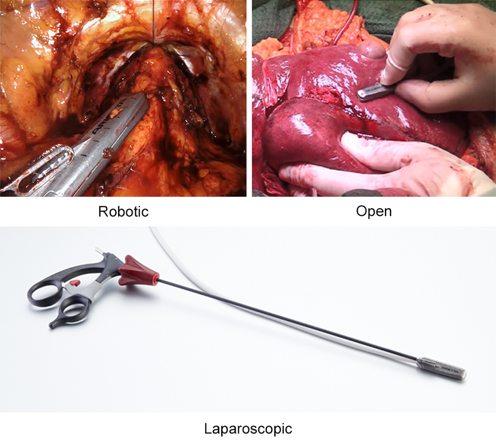
Three options – one probe for robotic, laparoscopic and open surgery
Every patient and every procedure is different. Which is why we have designed our drop-in probes to be versatile: you can firmly grasp the fin with a robotic surgery arm, use laparoscopic forceps instruments or your hand. Start minimizing cost and maximising functionality and choose your surgical approach every time: robot-assisted, laparoscopic and open surgery. With one single probe.
Ultrasound plays a key role in coloproctology for assisting the physicians prior, during and after a colorectal surgery. Combining high-end imaging technologies with our 360° electronic probe you get superior tools for accurate assessment and diagnosis of anus, rectum and colon pathologies with high confidence and great sensitivity. It helps planning the best therapy approach, and to follow-up the recovery process, enabling a safe and optimal patient’s outcome.
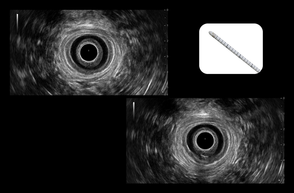
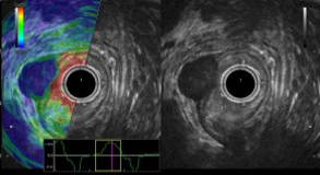
Assess from anal to rectal in a clear 360° radial view
Our electronic transrectal probe delivers high resolution radial ultrasound for clear localisation of even the smallest anomalies and assess a treatment approach. Get all anatomical details you need to diagnose pathologies, like fistulas, polyps or rectal lesions, by observing all different layers of the anal canal and rectal wall with the use of a balloon. In addition, the very slim probe – only 12mm in diameter – allows easy handling with less patient discomfort.
Get your best image for 360° Endo-Anal assessment
Scan in high resolution and detail the different anal canal portions (Lower, Mid and Distal) in order to identify sphincter defects, assess anal incontinence and the presence of abscesses. By checking the thickness and symmetry of the muscle and sphincter layers you can easily identify abnormalities, such as fistulas and follow their path.
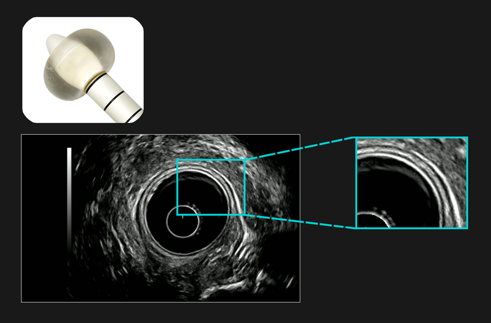
Get your best image for 360° Endo-Rectal assessment
To ensure a perfect coupling of the probe with the rectal wall, we use a balloon mounted on the probe head, inflated with water to have high-resolution images. Get a clear differentiation of the rectal wall layers (Mucosa, Sub-Mucosa, Muscularis and Serosa), which eases the process of rectal tumour staging. Our thin 12 mm diameter probe allows you to scan even in the presence of large rectal tumours, narrowing the rectal space, since it can pass into the rectal strictures.
Take a harder look at soft tissue to further characterize colorectal lesions
Improve cancer detection and staging with elastography, displaying differences in tissue elasticity in a colour map. The strain ratio index will help you differentiate between benign and malignant masses, while its real-time application facilitates to understand the extent of the tumour invasion. You can include assessment of lymph nodes, GIST or stage rectal tumours and decide on your treatment approach with confidence.
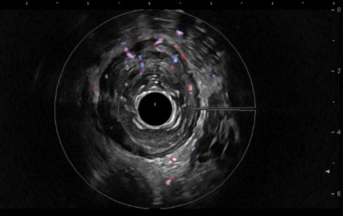
Understand vascularisation to outline suspicious areas
Dense or asymmetrical vascularisation in the rectal wall can be a suspicious sign for a cancerous lesion or reactive lymph node. Our highly-sensitive Colour Flow, eFLOW and Power Doppler modes display blood flow in greater detail – helping you identify areas of increased blood perfusion quickly and easily.
Ultrasound-guided blockage of peripheral nerves has refined many techniques in the routine practice of anaesthesiologists. From finding optimal vascular access to guidance of the needle to the target nerve and monitoring patients with TEE probes – our high-resolution and easy-to-use ultrasound solutions are your quick and efficient partner to help make surgeries safer.
Selectively block regional nerves with crystal-clear needle guidance
Navigate the local anesthetic directly to the target nerve by guiding your needle with our high-quality ultrasound imaging. Observing the surrounding anatomy and blood vessels in detail will help you to avoid damaging other structures on your needle pathway. And with our needle emphasis technology, you always know exactly where you are – and target even the smallest sensory branch nerves.
Control your patient’s status with transesophageal echocardiography
The highest image quality and hemodynamic analytics of our TEE probe help you notice physiologic changes during a surgery early, so you can react in a timely manner. In addition, you can monitor blood flow in detail with highly sensitive Doppler and Colour modes. This is especially beneficial in patients with a known cardiovascular disease who need diligent attention when undergoing anaesthesia.
Lightweight ultrasound probes, all day
Whatever you need, our family of highly functional probes can deliver it – from quality 2D cardiac images to 3D data; and from fetal heart assessment to pediatric and adult screenings. These extremely lightweight devices fit perfectly into your hand – for fatigue-free scanning, all day.
Choose the right solution for you
We know that no two clinics are the same. Your specific needs require a tailored solution: clinically, financially and ergonomically. Which is why we’ll work with you to find the ideal fit for your facility – from entry-level to premium advanced technologies.