Cardiovascular
Cardiovascular diseases count for one third of all global deaths. Are you ready to detect them early enough?
- Reduce the risk of missed diagnosis with superior ultrasound image quality.
- Detect early signs of heart failure with our hemodynamic analytics package.
- Get quick and consistent measurements with Cognitive Techonology-supported automisation.
- Diagnose structural heart disease using 3D/4D visualisations.
- Benefit from highly specialised and unique quantification tools for vascular assessment.
- Examine all generations – from fetal to pediatric to adult – using lightweight ultrasound probes.
- Achieve all this with solutions tailored to your unique needs, from premium to economy.
Get to your next level in cardiovascular imaging – with extensive analytics for the heart and vessels.
Our promise: a pure image
Above all, our cardiovascular solutions for ultrasound are based on excellent image quality. The interplay of image processing, probe technology and superior image optimization help capture the subtlest of signals. Which means you can see the cardiac and vascular anatomy in detail – for a confident diagnosis.
Switch from manual to automatic focus adjustment for homogenous imaging across all your patients
One common problem when applying Colour Doppler modes is the drop in frame rate. Frames get missed as the system struggles to follow the quick movement of the heart and images may not accurately mirror the actual blood flow. Our innovative Colour Doppler solves all this by maintaining a high frame rate together with a high resolution. So you can be sure not to miss a suspicious sign when evaluating blood flow.
Take it to the edge with Carving Imaging
Clear and sharp images are the key to a reliable cardiac scan. Our premium Carving technology pinpoints even the lightest echoes to enhance organ boundaries. You’ll get crystal-clear views of the myocardial borders, apical wall or valves; and you can assess even detailed structures with extreme accuracy.
Keep a high frame in Colour Doppler imaging
A common problem when applying Colour Doppler is the drop in frame rate. Frames get missed as the system struggles to follow the quick movements of the heart and images may not accurately mirror the actual blood flow. But our innovative Colour Doppler can maintain a high frame rate together with a high resolution. So you can be sure not to miss a suspicious sign when evaluating blood flow.
One solution to care for all patients – from fetal to children, to the elderly
Whatever your cardiovascular specialty, our ultrasound solution puts the right tool in your hand. You can combine the highest image quality, cutting-edge technologies and single crystal probes to care for patients of any age: from fetal to neonatal, from paediatric to adults and the elderly. We also offer tools for all ages: for example our TTE probes are ideal for patients from two to one hundred years old.
Solutions for everyone: from the fetus to senior patients
Heart defects affect patients of all ages, from the fetus to the 100-year old senior. While each have their own challenges in detection and treatment, the one critical requirement is image quality. Which is why our premium ultrasound solutions support the assessment of cardiac function throughout your patient’s life. One system – for clear images every time.
Identify congenital heart diseases earlier with our highly specialised fetal heart package
CHD in infants is a challenging issue, with structural cardiac anomalies often missed by prenatal ultrasound. To help improve detection rate, we offer you a sophisticated fetal heart imaging package that goes beyond routine screening. It gives you clear visibility and the valuable insights you need to make informed decisions in advising and treating your patient. Check here for more details!
Fast examination and clear images for neonatal patients
Thorough assessment of paediatric patients to avoid major problems in adulthood
If a cardiac anomaly is detected in a child, diligent monitoring and treatment is a must. But what about asymptomatic patients? Pathologies can develop as a child grows up – and access to detailed images of the coronary can be vital in its detection and treatment. Our highly sensitive Doppler and Colour modes depict even the smallest flows; while our new Mini TEE probe supports the planning and monitoring of heart surgeries.
Overcome echocardiographic disadvantages of high-risk elderly patients
The older a person becomes, the more difficult it can become to achieve high image quality. For example, patients might not have the endurance for a longer examination; or present a narrow cardiac window – despite being at high-risk of heart failure. Our highly sensitive ultrasound technologies, small probes and hemodynamic analytics respond to these challenges with ease: giving you crystal-clear images that differentiate between pathological changes and those associated with normal ageing; plus automated measurements for a fast examination.
Hemodynamic analytics
Heart failure is a prevalent medical condition and often comes along with hospitalization – impacting quality of life as well as healthcare costs. Now you can detect very early signs of heart disease with information gained from our hemodynamic analytics. This specialised package visualises and assesses cardiac blood flow from various angles, so you can base your decisions on grounded, clinical data – for accurate prognosis and patient treatment.
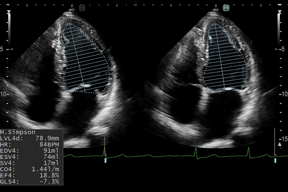
Measure the systolic function even in difficult patients with virtual contrast
Delineate the endocardial border of the left ventricle by simply applying our high-definition blood flow mode LVeFLOW. By overcoming wall motion noise, this highly sensitive technology delivers clear images of the fully filled LV cavity – even in older patients or those with a high BMI. You can use LVeFLOW instead of contrast agent imaging for a non-invasive and quick examination – with results that are comparable to MRI.
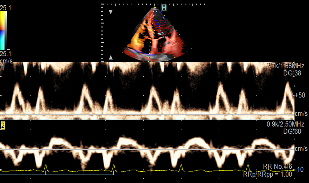
Get stable Doppler measurements even in patients with arrythmia
Our unique Dual Gate Doppler measures two separate sample waves from the same heart cycle at the same time. You can combine PW/TDI, PW/PW or TDI/TDI simultaneously, and improve your understanding of the diastolic function especially in patients with arrhythmia like atrial fibrillation. Using data from the same cycle makes the measurements very reliable, and calculates the E/e’, IVRT or IVCT indices extremely accurately.
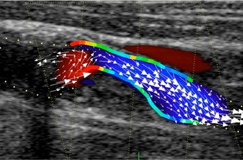
Visualise the hemodynamics within left ventricle with velocity vectors
Move beyond Colour Doppler imaging, where blood flow directions simply go up and down. Our innovative Vector Flow Mapping visualises flow dynamics in the heart and vessels in all directions as velocity vectors. So now you can view blood vortices in the cavity and check their formations, size and intensity. The result? Reliable data calculations help you detect suspicious signs of heart disease early – improving your prognosis, treatment decisions or follow-up.
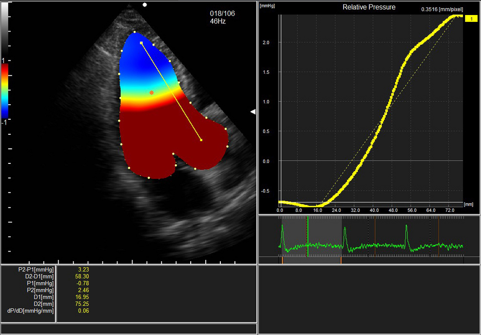
Non-invasive measurement of the relative pressure in the LV cavity
Most clinics use a catheter to measure the pressure in the left-ventricular cavity. But wouldn’t you prefer a non-invasive procedure? With our VFM, you can calculate the relative pressure gradient simply by conducting an echoscan. Just draw a line and click a button to get various pressure indices, like mmHg. The suction function in the isometric relaxation period is displayed in a pressure colour map – all of which enables you to assess diastolic function and quickly evaluate the heart failure risk.
Assess energy loss at turbulences to check treatment response
Turbulent blood flows in the left ventricle can be caused by stenosis or complications after surgery. Our VFM detects and characterises these flows by assessing the energy loss they cause: the more energy lost, the higher the turbulence. Studying energy loss – for example in patients with aortic insufficiency, or as follow-up before and after mitral valve treatment – has also proved useful in understanding hemodynamics.
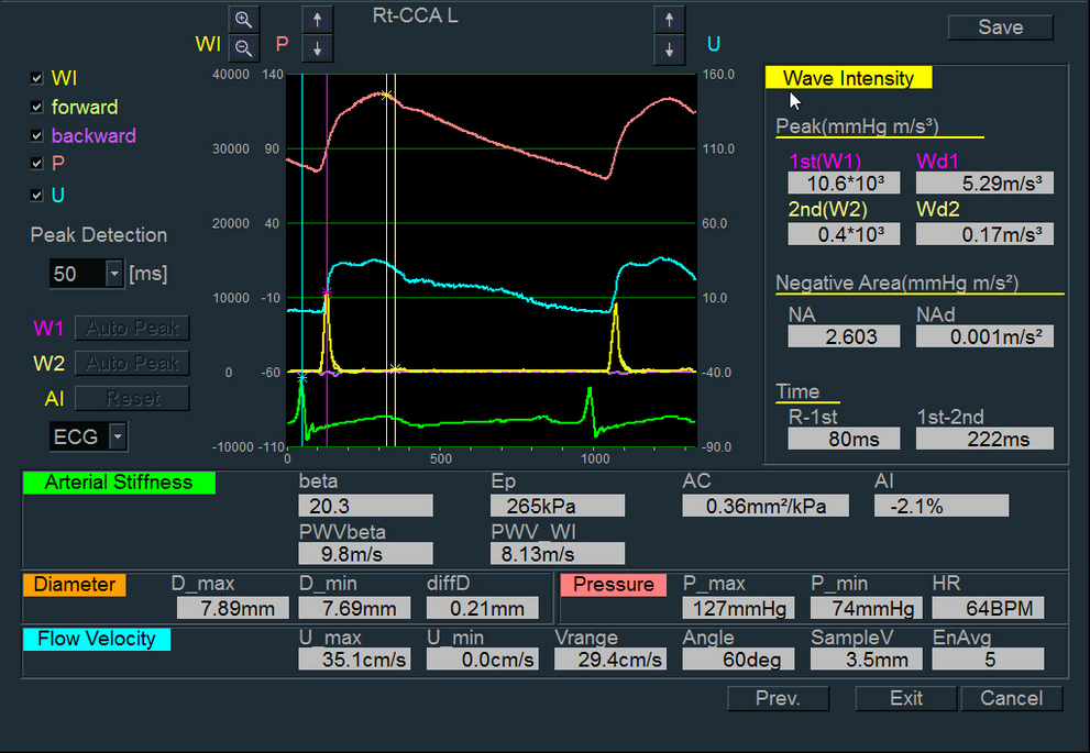
Add vessel analysis to your cardiac examination to get the bigger picture
The signs of heart failure aren’t limited to the heart. They can also appear in the vessels. The cardiac and the arterial system constantly interact with each other through waves that travel forwards and reflect backwards. Our unique Wave Intensity tool uses these travelling parameters – generated from vessel wall movements and velocity – to quantify circulation dynamics. Simply set your probe to the carotid artery to get insights into cardiac function – and even brain stroke risk.
Automated measurements for quick and accurate data
Automated detection of optimal frames for Ejection Fraction
Now you can drastically reduce your process steps when calculating the Ejection Fraction index. Our AI technology automatically recognises the optimal diastolic and systolic frames, and measures the EF based on the Simpson method. No more manual clicking through the frames and choosing the most suitable – because the system does it for you: quickly and accurately.
Cognitive Technology detection of stable R-R intervals for Doppler
Manually selecting stable R-R intervals can be challenging, especially if your patient has arrhythmia. Adding another manual measurement of the E/e’ index means that you end up spending lots of time simply operating the system. The good news: now you can do it in a single click! Our iDual Gate Doppler automatically detects stable R-R intervals, measures PW/TDI in the same cycle, and delivers E/e’ in just five seconds.
Quantify and analyse the complete myocardial function in just one click
Add comprehensive clinical information with our high-power 3D/4D package and high-resolution 3DTEE and 3DTTE probes. Realistic ultrasound images help you look closely at cardiac anatomy and detect even small anomalies. Then you can analyse the data later on and conduct more detailed measurements for structural heart disease and valvular pathology cases.
Understand structural anomalies with 3D and bi plane views
Not only can you improve the visualisation of valve structures or the thrombus with our high resolution, realistic 3D/4D images. You can then use our 3DTEE and 3DTTE transducers to create high quality 2D images for clearly assessing the anatomy in the bi plane view. Access the 3D volume reconstruction offline after the examination to analyse the details for a confident diagnosis and process planning of your treatment.
Quantify mitral valve anatomy to guide treatment decisions
For complex mitral valve pathologies like a prolapse, you need a detailed assessment to decide and prepare the right therapy. With 4D MV Assessment*, you can analyse and quantify its anatomy and movement. Set reference points on the 3DTEE image and automatically obtain important geometric measurements like annular dimensions, leaflet morphology or coaptation descriptions. Then use this data to help plan your patient’s therapy and monitor the treatment response.
Visualise and quantify the full LV function in 3DTTE
Use 4D LV-ANALYSIS* to assess the left ventricular function based on 3D speckle tracking. This innovative research approach displays the regional and global strain analysis via a 3D model of the LV that visualises the wall motions comprehensively; while a colour-coded bullseye highlights healthy and problematic areas in different segments. In all, it gives you a better understanding of wall motion anomaly and cardiac function.
Avoid MRI for RV function assessment with 3DTTE
The right ventricle can be difficult to assess in 2D ultrasound because of its position which is why most patients get referred for MRI. However, recent studies have shown that 3D echocardiography delivers similar results to MRI and it’s quicker and easier. 4D RV FUNCTION* analyses and quantifies RV function for cases like pulmonary hypertension or right sided heart failure; and it enables you to combine 3D and 2D values including EDV, EDVi , ESV, ESVi , EF and SV, RVLS, TAPSE and FAC all from the same volume data set.
* Module from TomTec-Arena™, a registered trademark of TomTec Imaging Systems GmbH in Germany and the United States of America.
Early detection and prevention of cardiovascular diseases like diabetes or arteriosclerosis is especially essential because they can affect other organs as the blood travels through them. It’s why we developed specialised tools for the morphological and functional assessment of vessels – giving you reliable data for a confident diagnosis.
One probe to view all vessels with our unique CMUT
It can take up to three probes to view vessels at different depths. So why not save yourself some trouble and choose our CMUT linear probe. It visualises all vessels with ease; and the frequency bandwidth from 22 – 2 MHz can penetrate near and deep anatomy identifying even small vessels throughout. You’ll get clear, high resolution images in B mode, Colour and Doppler modes. So why wait? Start saving time and improve your imaging right now.
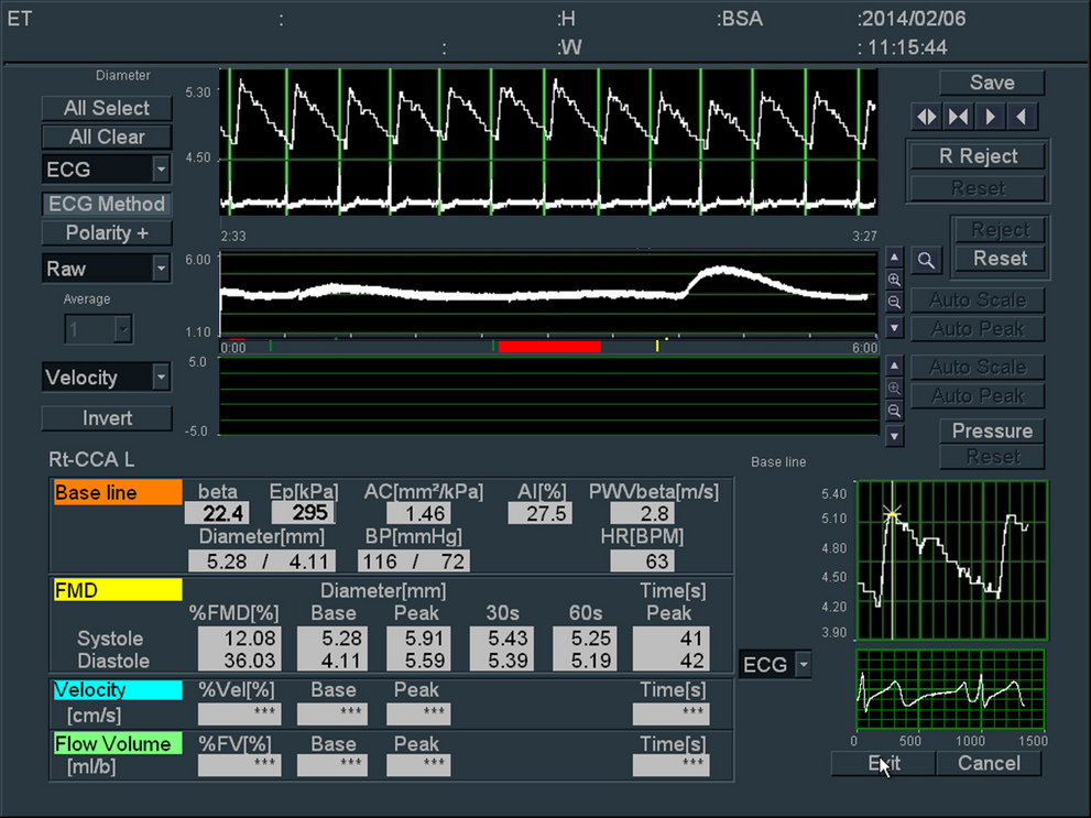
Assess the risk of arteriosclerosis with Flow Mediated Dilation
A very early sign that your patient might develop arteriosclerosis can be found in the endothelial function of the vessels. With our FMD, you can measure the changes in vessel dilatation when blood flow volume is increased. The resulting parameters such as %FMD or peak velocity give you detailed insights into the vessel wall condition. All of which helps you take the right preventive steps and improve patient care.
Evaluate the progression of early arteriosclerosis via arterial stiffness
Hypertension can be an early indicator of arteriosclerosis. Now you can confirm this disease with eTRACKING checking the vessel wall stiffness on the carotid. Compared to an ABI test, it’s quick, stable and unique. We are the only vendor offering this detailed level of insights. In fact, you can automatically obtain a comprehensive range of stiffness parameters like ß, PWV, Ep and others within seconds. For more on this topic check out this recently published study on European normal values by age and gender.
Combine plaque and velocity data to manage arteriostenosis
Plaque can cause severe arteriostenosis, and patients may need different care depending on type and grade. Now you can characterise plaque in detail with our high resolution B mode and check its vascularity with the highly sensitive DFI. Combine this information with Continuous Wave Doppler using the same probe to quantify velocities in the blood flow around it. You can obtain a detailed overview of the plaque and its risk of reaching the brain and make confident treatment decisions.
Assess the vascular system in the whole body of your diabetes patients
We know you need to understand and monitor the whole vascular system for patients with certain vascular diseases like diabetes – which can develop into atherosclerosis or stenosis in peripheral vessels. Which is why our ultrasound solutions help you view the endothelial function, vessel wall thickness and stiffness, plaque or blood flow velocities in detail. As an additional research tool, our Vascular VFM can even quantify wall shear stress to assess the possibility of vascular access.
Increase efficiency and accuracy with Cognitive Techonology based automatisation
Ultrasound-guided blockage of peripheral nerves has refined many techniques in the routine practice of anaesthesiologists. From finding optimal vascular access to guidance of the needle to the target nerve and monitoring patients with TEE probes – our high-resolution and easy-to-use ultrasound solutions are your quick and efficient partner to help make surgeries safer.
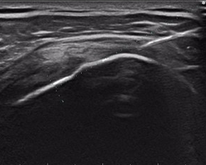
Selectively block regional nerves with crystal-clear needle guidance
Navigate the local anesthetic directly to the target nerve by guiding your needle with our high-quality ultrasound imaging. Observing the surrounding anatomy and blood vessels in detail will help you to avoid damaging other structures on your needle pathway. And with our needle emphasis technology, you always know exactly where you are – and target even the smallest sensory branch nerves.
Control your patient’s status with transesophageal echocardiography
The highest image quality and hemodynamic analytics of our TEE probe help you notice physiologic changes during a surgery early, so you can react in a timely manner. In addition, you can monitor blood flow in detail with highly sensitive Doppler and Colour modes. This is especially beneficial in patients with a known cardiovascular disease who need diligent attention when undergoing anaesthesia.
Choose the right
solution for you
We know that no two clinics are the same. Your specific needs require a tailored solution: clinically, financially and ergonomically. Which is why we’ll work with you to find the ideal fit for your facility – from entry-level to premium advanced technologies.
Lightweight ultrasound probes, all day
Whatever you need, our family of highly functional probes can deliver it – from quality 2D cardiac images to 3D data; and from fetal heart assessment to pediatric and adult screenings. These extremely lightweight devices fit perfectly into your hand – for fatigue-free scanning, all day.


Hear from others
High quality cardiac imaging in critical patient care
Prof. Paolo Trambaiolo, intensive cardiac care in Rome
High quality cardiac imaging with LISENDO 880
Dr. Dewyani Chowdhury, paediatric cardiologist in Pennsylvania