
Women's Health


See details
See details
See details
See details

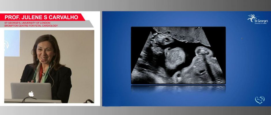
Advanced fetal heart examinations and cardiac function assessment
Lecture by Prof. Dr. Julene Carvalho
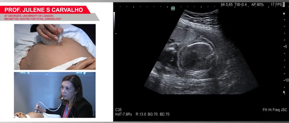
Fetal heart examination and cardiac function assessment
Lecture by Prof. Dr. Julene Carvalho
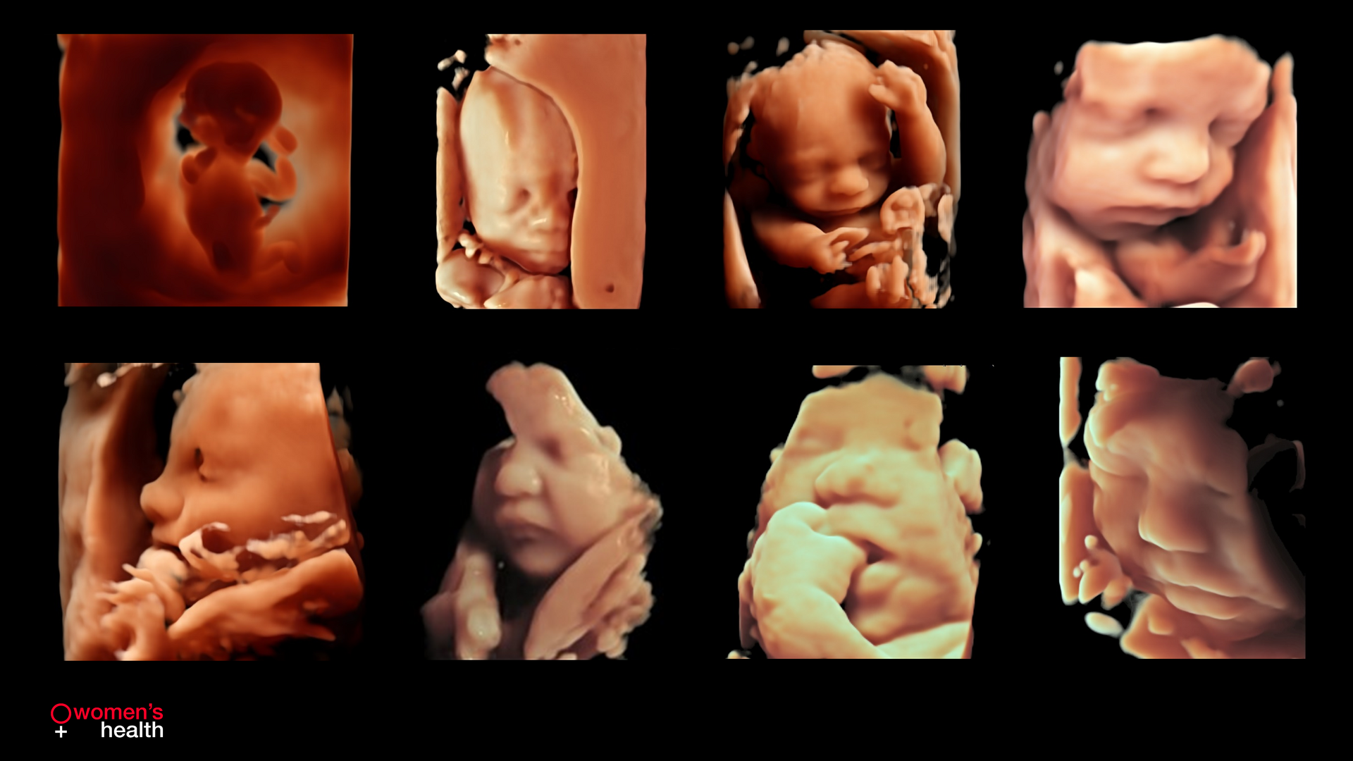
Detailed 3D/4D ultrasound visualizations from early to late pregnancy. We offer sophisticated tools like 4Dshading, Stagelights and Specular to create realistic images.
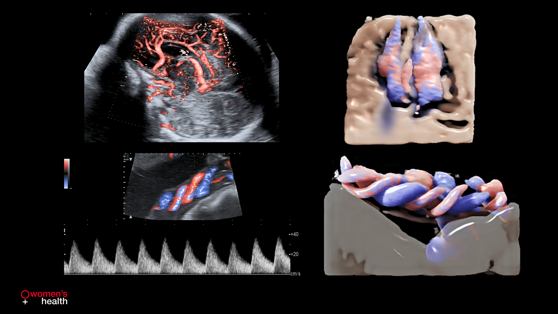
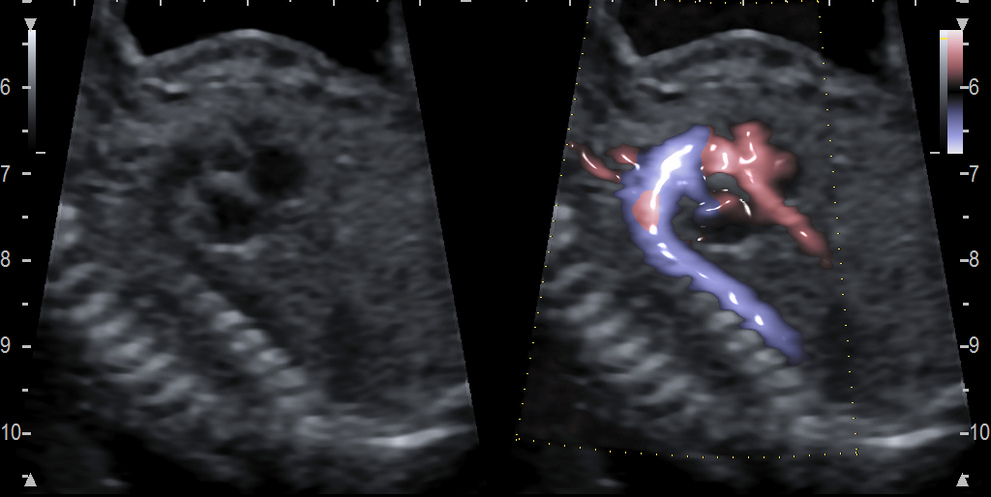
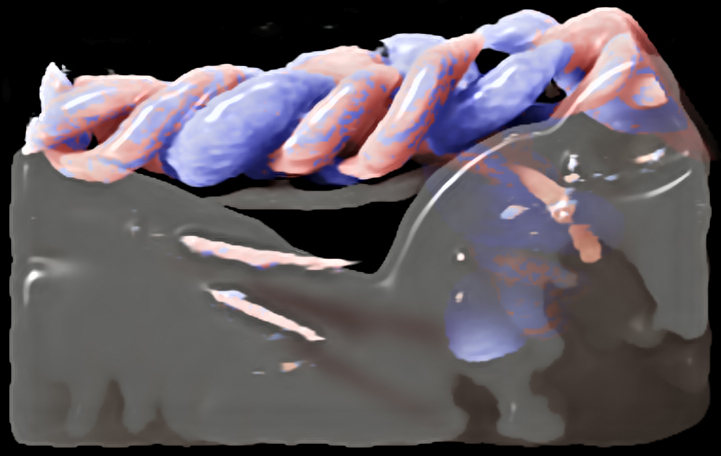
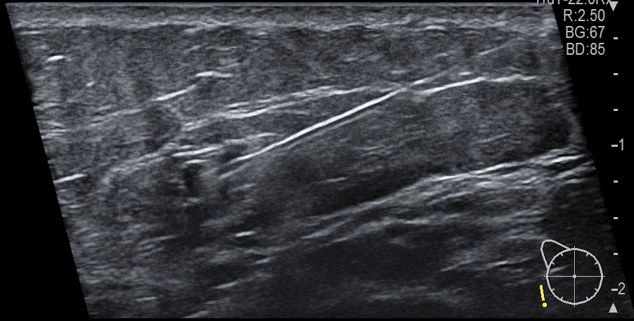
We offer various vascular imaging solutions to depict and assess vessels in great detail, like Flow, Power Flow, eFLOW, DFI, Glossy mode, Dual Gate Doppler and 4Dshading Flow.
To help improve detection rate of CHD, we offer a sophisticated fetal heart imaging package that goes beyond routine screening, with high-resolution B-mode images, Glossy mode, Dual Gate Doppler, STIC or 4Dshading Flow.
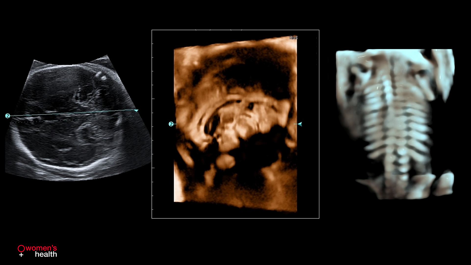
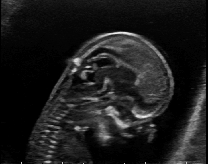
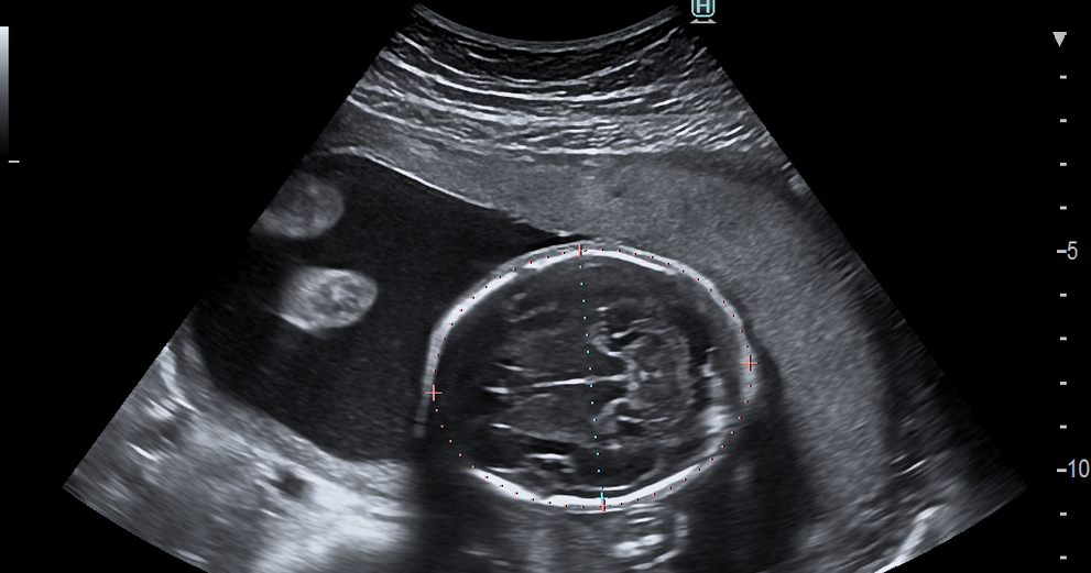
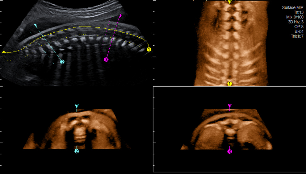
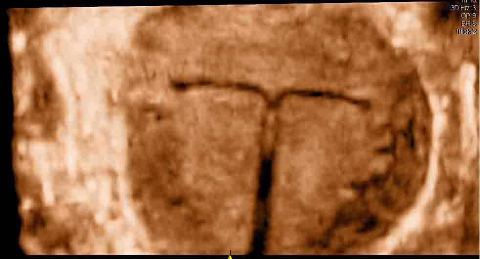
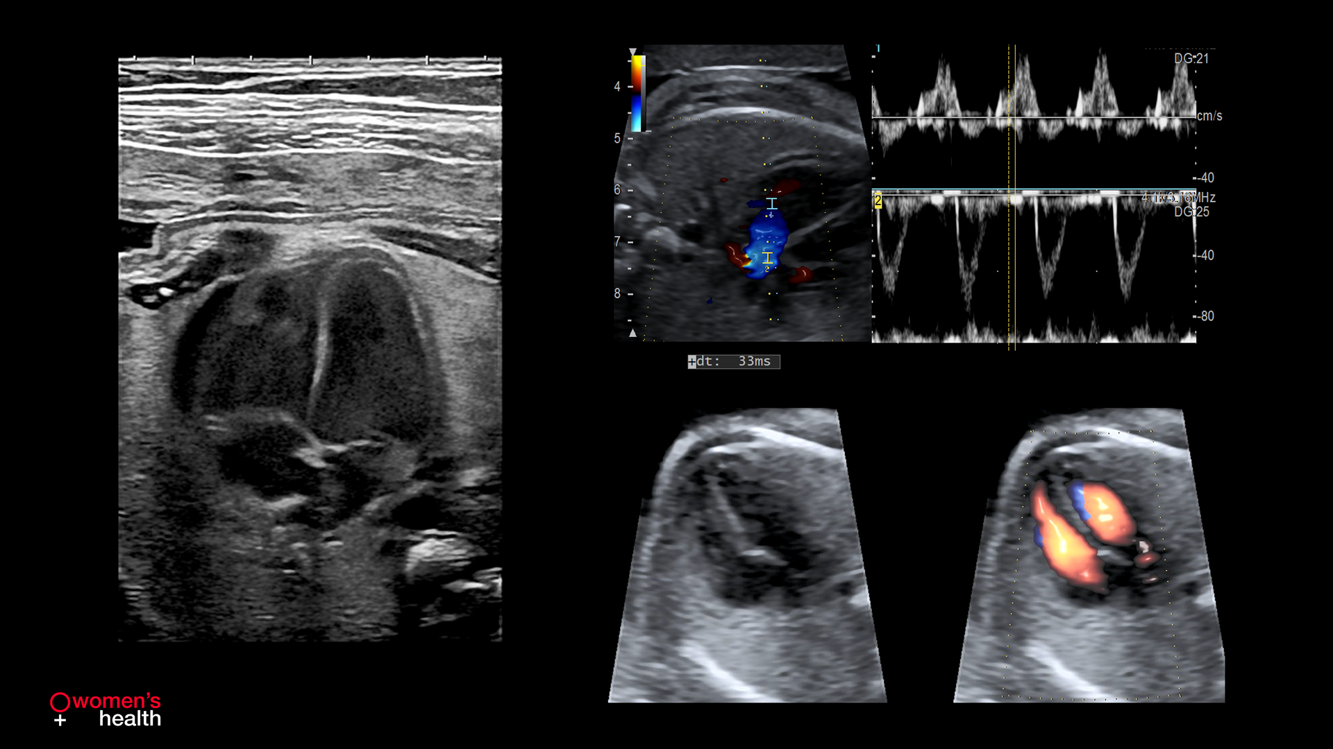
Clear ultrasound visualizations of the central nervous system (CNS) in high-resolution B-mode with dedicated tools to improve diagnostic confidence, such as CMPR and 3D/4D renderings.
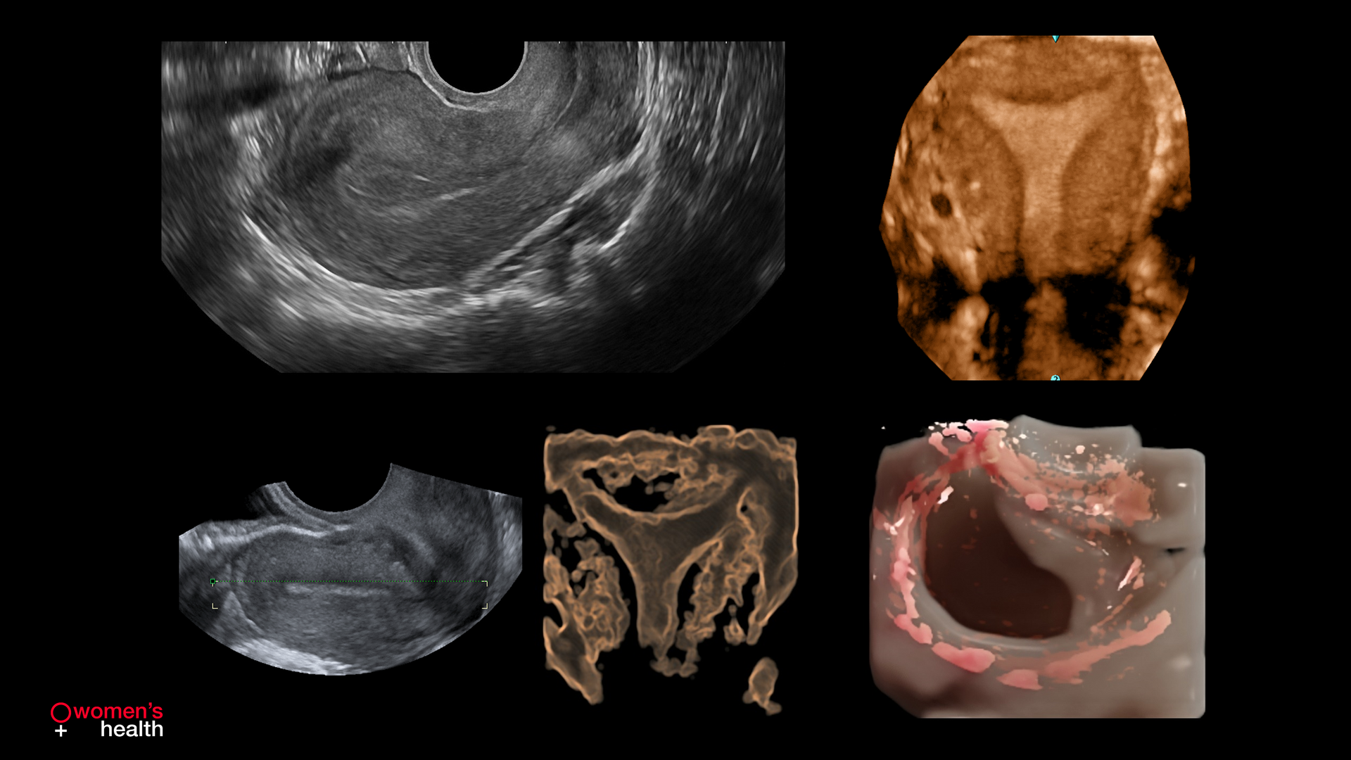
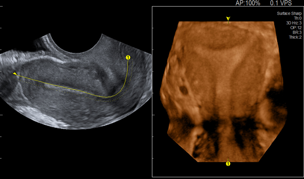
Ultrasound of uterus and ovary using high resolution B-mode, 4Dshading or CMPR to see it in the curved coronal view for detailed assessment.


