
Urology
Urology. You expect quick, precise and efficient support from ultrasound imaging.
So that’s what we deliver. Our flexible ultrasound solutions support doctors to diagnose a wide range of clinical conditions with different treatment strategies – from a standard screening process to highly specialised interventional procedures.
- No matter what your approach is – we have the right ultrasound transducer for you.
- Get a clear picture with our superior ultrasound image quality.
- Enrich your procedures with advanced features like fusion and contrast imaging.
- Biopsy the prostate confidently – from transrectal or transperineal, to systematic or targeted.
- Manage kidney pathologies by looking at vascularisation in detail, and perform precise treatment guidance.
- Find sources of pain, swelling or infertility in the testes and penis quickly and accurately.
- Perform minimally invasive urological surgeries with precise ultrasound tools.

Detect, biopsy, diagnose, treat and follow up - with high-quality ultrasound solutions for all stages of urology patient care.
Prostate Imaging
Despite prostate cancer being the most common cancer among European men, we know that patients can be reluctant to see the doctor. This is why our ultrasound solutions are designed to make this process as fast and precise as possible by helping you screen, assess, biopsy and treat your patients more efficiently. Choose from a wide variety of probes and features to match the best biopsy approach – from transrectal or transperineal, to systematic, freehand or targeted.
Choose the transducer that fits your approach
See suspicious signs early and accurately to locate the right spot
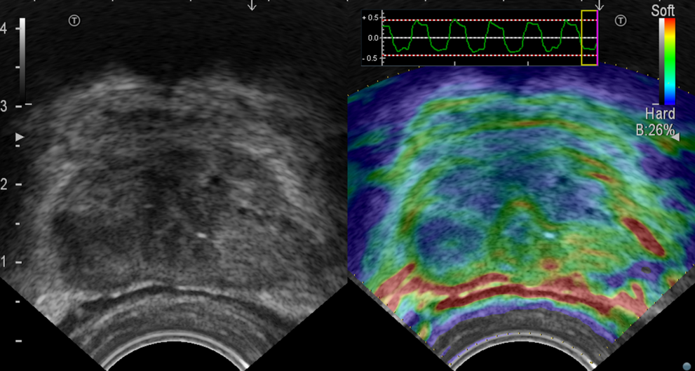
Take a harder look at soft tissue to distinguish affected from healthy areas
Understand vascularisation within the prostate to identify suspicious areas
Dense or asymmetrical vascularisation in the prostate can be a suspicious sign for a cancerous lesion because it’s nourished by the nearby blood vessels. Our Colour Flow, eFLOW and Power Doppler modes display blood flow with high sensitivity – helping you identify areas of increased blood perfusion quickly and easily.
Get a close look at microvascular perfusion
Perform transperineal prostate biopsies under local anesthesia
Plan and guide prostate biopsies with MRI-US fusion imaging
Get a significant tissue sample the first time with an accurate biopsy guidance – whether you approach transperineally or transrectally. With our embedded fusion imaging solution you can combine prior mpMRI data with the ease-of-use of real-time ultrasound guidance. You can even add various ultrasound imaging modes for even greater precision. In fact, these types of targeted biopsies are now highlighted in the recent update of the EAU prostate cancer guidelines.
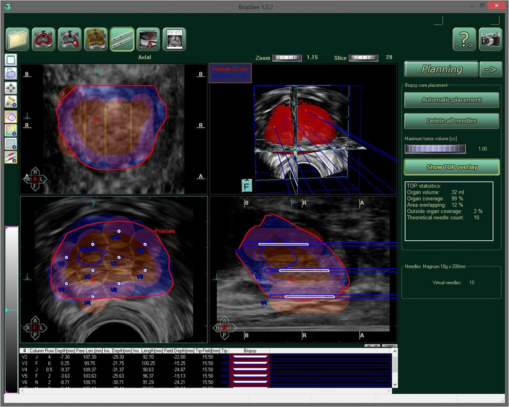
Combined forces for prostate biopsies and therapy
Performing procedures like a transperineal prostate biopsy takes some practice. For practitioners who prefer more dedicated support, we’ve therefore teamed up with BiopSee by MedCom. This prostate-dedicated MRI fusion solution facilitates the process from pre-planning to real-time guidance instructions and core registration – delivering extensive reporting or 3D reconstruction. Similarly, it also provides extra assistance and guidance during focal therapy treatments.
Minimally invasive procedures for brachytherapy or focal therapy treatment
How can you target prostate cancer therapy more precisely? Increasing the accuracy of brachytherapy, cryoablation or electroporation will destroy cancer cells while avoiding damage to healthy tissue; while innovative approaches also protect rectal tissue by injecting hydrogel. For this kind of precise needle guidance, we offer biplane probes to investigate the prostate in both the transverse and longitudinal planes. By combining this with high-quality, complementary imaging modes, you can be sure to hit your target.
Kidney & Bladder Imaging
Check the anatomy and functionality of the urinary system with high-quality ultrasound solutions. For disease management, we offer advanced features for precise biopsy and treatment procedures – supporting you through all stages of patient care: from detection to diagnosis, treatment and follow-up.
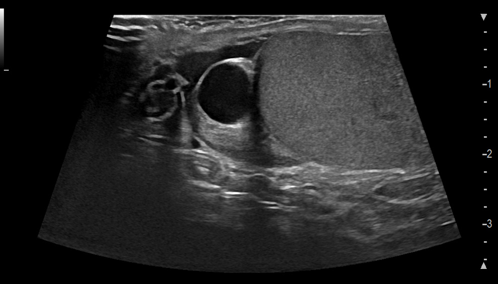
Get clear tissue differentiation – whatever the BMI of your patient is
In transabdominal ultrasound it can be hard to find the balance between a good resolution and a good penetration – because every patient has a different body type. With our refined technologies there’s no need to compromise, thanks to crystal-clear boarder visualisation and tissue differentiation at any depth. Investigate the location, shape and inner structure of the renal anatomy, parenchyma or sinus in high resolution to screen for abnormalities like blockages, stones, tumours or cysts.
Assess blood flow into, out of and within the kidney with high sensitivity
Our highly sensitive Colour and Doppler modes help you gain a deeper understanding of renal perfusion and check for asymmetries or non-vascular areas. Furthermore, you can quickly assess the blood flow within lesions and their surrounding tissues to help differentiate between benign and malignant signs.
Get a close look at microvasculature for assessment, biopsy or treatment
Our contrast-enhanced ultrasound delivers vascular information beyond Doppler possibilities. You can view wash-in and wash-out times for circulatory information on the renal parenchyma and focal lesions. You can characterise hypovascular or atypical tumours; solid or cystic masses; pseudotumours; ischaemia; or infections. You can also use it for precise needle guidance during biopsies, radiofrequency, cryo or microwave ablations.
Plan and perform precise needle guidance with automatic CT-US fusion imaging
Kidney biopsies or tumour ablations may be challenging, but you can find a precise, safe needle path with our fusion imaging. Real-time ultrasound is automatically synchronised with prior CT images to help you locate the lesion accurately and plan the optimal access path and patient positioning. You can then guide the procedure with needle tracking and body motion compensation, without additional radiation exposure. At last, you can better monitor treatment outcome.
Leave no stone left unturned with our specialised kidney biopsy probe
Percutaneous nephrolithotomy (PCNL) is today’s standard for removing large or otherwise persistent kidney stones. Our biopsy ultrasound probe helps you precisely locate them and guide direct access. Simply insert the needle through a cut-out in the centre of the probe at three different puncture angles. With firm insertion control, you no longer need to navigate around the lateral rib position to find your way. Detach the probe from the needle after hitting its target in one single action. Watch this video to see it in action!
Check bladder condition and draining to find the source of a problem
Our high-resolution Carving Imaging enhances organ boundaries and highlights contrast resolution between fluids and tissues. Together with a sensitive Colour Doppler, you can easily investigate the bladder, its wall and volume or the ureteral jets. Findings such as pre-and-post void residual, wall diverticula, urethral stricture, stones or tumours will help determine the cause of the problem and guide you in providing treatment options.
Testes & Penile Imaging
Ultrasound scans can play a key role in assessing the male reproductive system, especially when the patient is in pain and needs immediate care. That’s why our sensitive B-mode, Colour, Doppler, elastography and contrast modes deliver high-resolution imaging for a fast and accurate assessment of swelling, inflammation, trauma or torsion – as well as lesions and problems with blood flow supply and infertility.
Detect and localise anomalies with greater speed and precision
We’ll help you get a clear delineation of structures for detecting anomalies like an undescended testicle or plaques; finding lesions; and assessing the integrity and thickness of the tunica albuginea. One invaluable tool here is our innovative CMUT linear probe – which incorporates the highest resolution, penetration and a super-wide frequency bandwidth with the newest ultrasound technology. So now you get the full picture in one glance, smoothly guiding your patient management.
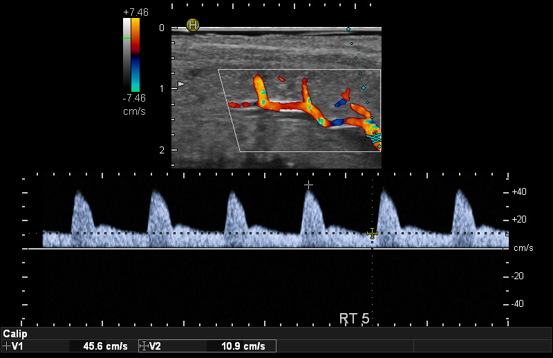
Assess blood supply to find sources of erectile dysfunction and infertility
Apply our highly sensitive Colour and Doppler modes to study the blood flow supply and diagnose erectile dysfunction. You can also detect inflammation or observe blood flow of a testicular torsion or a twisted spermatic cord. Our solutions help you assess vascularisation and blockages with ease – resulting in a confident patient consultation about possible treatment options.
Take a harder look at soft tissue for further differentiation
Malignant focal lesions tend to be stiffer than healthy tissue. Which is why back in 2003 we were the first company to launch a strain elastography solution. You can improve cancer detection and border delineation by displaying differences in tissue elasticity via a colour map. It’s quick, easy to understand and a standard feature in all our linear ultrasound probes.
Get a close look at intratumoural microvascularisation with CEUS
With our contrast-enhanced ultrasound you can understand vascularisation beyond Doppler possibilities – for example when imaging hypovascular or avascular lesions. It helps you assess more confidently – from testicular segmental infarctions, to the extent of viable tissue after trauma, to the feeding artery. Which means you’re quipped with more information to make improved clinical decisions.
Interventional procedures like nephrectomy or prostatectomy can really benefit from a minimally invasive ultrasound-guided approach – reducing patient trauma and accelerating recovery time. Which is why we offer dedicated robotic drop-in and laparoscopic probes with advanced, high-resolution imaging modes for real-time information – enabling you to operate safer, faster and with more flexibility.
Robot-assisted procedures with precise ultrasound guidance and full control
Our drop-in probes help robotic surgeons navigate inside the human body and make critical decisions, fast. The linear array provides high-resolution imaging and optimal organ; while a virtual convex mode enables an enlarged field of view. It all comes with a small footprint, flexible cable and lightweight design for excellent in-situ usability with a full-wrist, 360° articulation.
Laparoscopic ultrasound for minimally invasive surgeries
Improve patient outcome and cost-effeciency with our four-way flexible laparoscopic probe. Insert it through a trocar and see beyond the surface into the organ anatomy, locating lesions by differentiating between tissue structures with high-quality B-mode and elastography ultrasound. You can plan your procedure with Contrast imaging for clear resection margins; and avoid injuring important vessels during resection with our sensitive Doppler and Colour modes.
Reduce WIT in kidney surgery with contrast-enhanced ultrasound
Warm Ischemia Time (WIT) is a crucial factor in kidney surgeries. But to reduce the impact of lost blood supply, you can only clamp specific arteries during the procedure. Our contrast-enhanced ultrasound helps you by targeting these key arteries, down to capillary perfusion. This in turn helps you avoid global ischemia, thus reducing the permanent loss of nephrons.
Choose the right
solution for you
We know that no two clinics are the same. Your specific needs require a tailored solution: clinically, financially and ergonomically. Which is why we’ll work with you to find the ideal fit for your facility – from standard to specialised probes, from entry-level technology to premium advanced applications.

Hear from others
Moving premium features to high-end: Renal & Fusion imaging with ARIETTA 750
Lecture by Prof. Dr. Jean Michel Correas, Paris