Women's Healthcare Technologies

FUJIFILM is proud of the reputation we’ve built as an industry leader in Women’s Imaging. Known for our unparalleled image quality, superior system reliability and intuitive use of cutting edge technology, FUJIFILM remains the ideal choice for exceptional diagnostic ultrasound imaging in the field of Women’s Healthcare.

3D/4D Imaging Technologies:

Volume Slice Imaging (VSI)

Displaying cross-sectional images with thickness, VSI improves image quality by enhancing contrast resolution and reducing speckle noise, and enables easier 3D understanding of the target. The image is showing a detailed image of the fetal brain using VSI.

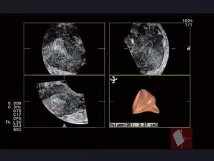
Multi-Follicle Volume (MFV)

Follicle growth management by follicle diameter is performed in many infertility treatment facilities today. However, it is not easy to consistently obtain accurate measurements, as follicles are soft and can appear in various shapes.
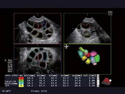
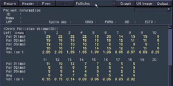
MFV automatically calculates the number and volume of areas with low brightness within the volume data. This function is effective in measuring volumes of multiple follicles and understanding of follicle numbers.


Example of a report screen. Measurement results are automatically transferred to the report, which can easily be displayed and printed out.

Morphological Evaluation and Volume Measurement using Volume Data
Volume data can be used for morphological evaluation and volume measurement of the uterus and ovaries, in addition to follicle evaluation.

Instantly get volume data of superficial areas
ASU-1013, the high frequency probe for scanning superficial areas in 3D, was expertly designed especially for breast examinations.

Its structure and materials contribute to clear imaging in both 2D and 3D with high penetration that reaches to as far as the greater pectoral muscles.

Easy Volume Data Acquisition

  • Volume Data can be acquired in a short scan. Once and image is acquired, 3D reconstruction can be done at any time.

  • The Check Scan function is useful for making sure the target area is in the scanning range.

  • Supports acquisition of Volume data in Power Flow Mode

Real-time Tissue Elastography (RTE)

Elastography with Strain Ratio Measurement designs an easy workflow to detect and evaluate masses in breasts, and is expected to increase the detection rate of breast cancer.

Elastography visualizes tissue stiffness in real time, and Strain Ratio Measurement will then display the ratio of deformation in two areas of your choice. The deformation ratio of fatty tissue and the lesion, or Fat Lesion Ratio (FLR), can be easily calculated, providing exam results quantitatively. Because what is calculated is the ratio, the acquired quantities are stable.