SCENARIA™ View
CT scanner
Superb low-dose CT
Getting the best image quality with the lowest dose – for every patient, every time – has always been a huge challenge for CT exams.
But not anymore. Our premium SCENARIA View CT scanner combines high image quality and low dose with no problem.
This solution not only delivers natural, clear image textures at lower dose levels than ever before; it also comes with the industry’s widest bore design – and an innovative automation package that improves your productivity and patient throughput.
- Iterative processing technology Intelli IPV enables low-dose exams with no compromise on image texture – ideal for all your clinical challenges.
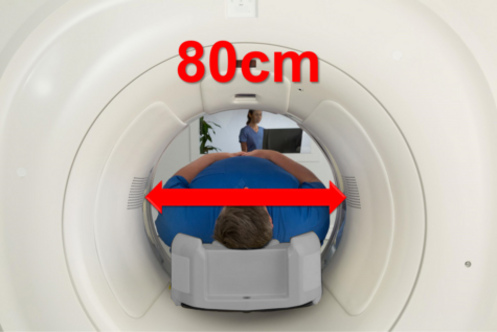
- The open design with an 80cm-wide bore eases patient anxiety.
- Places target regions in the FOV centre thanks to a patient table that slides 20cm to the left and right.
- Provides great patient access and positioning options – enabling interventional CT procedures.
- Enhances productivity and patient throughput with SynergyDrive Automation Suite, an automatisation suite for patient positioning, protocol selection, high-speed image calculations and post-reconstruction.
Why choose between low radiation dose and excellent image quality?
Now you can get both with SCENARIA View’s CT scanner iterative reconstruction technology Intelli IPV. Visual Modelling reduces dose and noise with no compromise on image texture. And our dose control tools help you adapt your exams to the precise needs of each patient.
- Automatically modulate mA to lower dose levels for each patient depending on their anatomy and size – plus the level of iterative reconstruction selected – with INTELLI EC PLUS.
- Small bowtie filter precisely targets radiation only where needed.
- Higher data sampling rate provides faster anatomic coverage without sacrificing image quality, while reducing motion artefacts.
- Lower kV settings to scan below the typical 120 kV – ideal for paediatric patients or for better contrast detection.
- Reduced mA dose for cardiac CTA during the cardiac phases of a retrospectively gated exam.
- Dose Reporting complies with all legal and clinical documentation requirements.
It all starts with patient comfort.
The more relaxed your patients are, the clearer their images – which leads to more accurate diagnosis and treatment decisions. That’s the simple philosophy behind SCENARIA View CT scanner: the ideal blend of clinical functionality and patient comfort.
- Industry-leading 80cm wide bore eases patient anxiety and accommodates obese patients and children.
- Allows flexibility of scan positions thanks to a spacious design.
- Enables patients who struggle to raise their arms to be scanned with their arms by their sides (like the obese, elderly or trauma patients).
- Puts target regions in FOV iso-centre for the best resolution thanks to a patient table that slides 20cm left and right.
- Suitable for patients up to 250 kg.
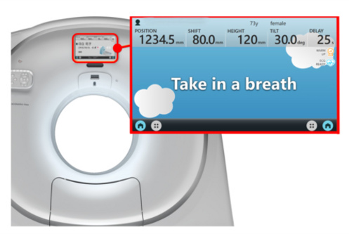
- Central control panel manages all operations of the table and the gantry.
- Patient instructions on 15” touch monitor in various languages, including a version for children.
Streamlined workflow for high-speed exams
Technical functions supporting your clinical work
- Correct image artefacts caused by metal implants using HiMAR Plus.
- Move the table back and forth for complete blood flow images in the brain (extended coverage for perfusion exams) with Shuttle scan.
- Dual Energy with double rotation method enables analysis of material composition through acquisition at two different kV energy levels.
- AutoPose with completely automatic scan range recognition – without the technician touching the mouse.
Special features for specialised needs
- Visualise organs from the inside – like the colon, bronchi, stomach, blood vessels, bladder, kidney, larynx or paranasal sinuses – through Cruising Eye View (CEV-CPR) with virtual endoscopy.
- Predict scan with bolus tracking for automatic scan start once the contrast agent has reached target vessels.
- Quantify blood flow through the brain parenchyma with Perfusion Stroke Analysis.
- Perform a complete biopsy or pain therapy in real time from a large monitor in the scanner room Guide Shot for CT fluoroscopy.
- Enhanced Cardiac Package with effective coronary CT angiography (CTA) and calcium scoring tools.
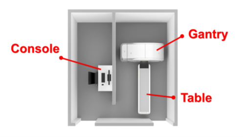
SCENARIA View CT scanner is a true three-module system: consisting of just a gantry, table and console. No transformers or separate units are needed. It also comes with a powerful engine, reconstructing up to 128 slices per scan rotation – with quality images for a confident diagnosis.
- Max. scan speed of 0.35 seconds for routine, high-speed protocol scanning.
- 40mm detector coverage.
- 64 discrete detector row & electronics, with 128 slice Fine Recon.
- X-ray tube: 45 MHU equivalent.
- Generator: 72kW standard, 84kW optional.
- Easy siting with only three modules (gantry, table, console).
- EcoMode reduces standby power consumption by up to 70%.
- Remote application support to help optimise protocols or train technicians online with SENTINEL™.
- Remote maintenance support delivers automatic notification of anything suspicious via SENTINEL™ – maximising your uptime.