RADAR
Clear images minus the motion artefacts
Wouldn’t it be great if your patients always laid as still as a stone?
Unfortunately, the reality is that they move around during their MRI examination – whether due to anxiety or natural reflexes, like heartbeat, breathing and swallowing, or both.
MRI has always been sensitive to motion artefacts. And the longer a sequence takes, the greater the chances are of them degrading the image quality with smearing, blurring or ghosting. Confusing such artefacts with a pathology can lead to serious misinterpretation. So, to get clear images with little motion artefacts, radiologists may even have to sedate their patients.
But it doesn’t have to be this way…
Now you can make up for body movement during MRI exams with our industry-leading RADAR motion compensation technique. Suppress unwanted signals at the touch of a button. And you don’t need to compromise on image quality, scan time or application: RADAR is compatible with any coil for any anatomy – and most sequences, including:
- Fast Spin Echo for morphological imaging in any anatomy.
- Fast Inversion Recovery to suppress fat or CSF signal.
- Spin Echo for real pure T1 contrast.
- 3D TOF angiography for vascular applications.
- T2* Gradient Echo for neuro or orthopedic studies.
- Fat suppressed real T1 post contrast.
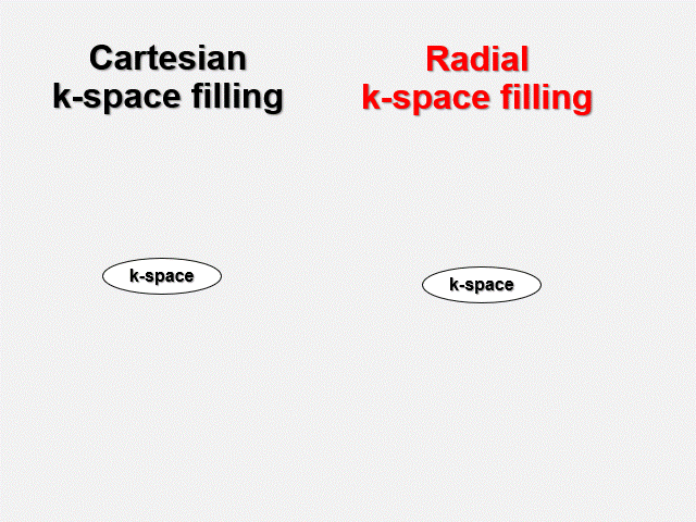
Overlay radial scans in the centre of the k-space.
Cartesian sequences are the most common encoding method in MRI – and they’re very sensitive to patient motion. RADAR follows the RADIAL ACQUISTION REGIME, where each blade crosses the k-space centre, overlaying image-by-image. The result: better SNR, CNR, and significantly less motion artefacts. Which is precisely why RADAR is the industry-leading technique in its class.
Conduct a complete brain studyminus the motion artefacts with All Around RADAR
Patients often suffer pain and confusion when undergoing brain MRI scans due to trauma or strokes. Meanwhile, children are often scanned with MRI rather than CT to avoid radiation. In both cases it can be hard to persuade patients to follow instructions and lay still. Which is where our All Around RADAR can help you deliver a solid, reliable and fast solution for all sequences needed for a complete brain study – including DWI, TOF, GE or T2*.
Triple Combo for motion-free, fast and silent MRI exams that relax your patient
Our SoftSound technology combines software and hardware features for quiet scans that meet your diagnostic needs. What’s more, you can combine it with RADAR, for motion artefact reduction, and RAPID, for high-speed imaging.
The result? A fast, silent and motion-free MRI exam: for every coil, anatomy – and patient.
