Low MI contrast specific imaging technology developed for use in conjunction with ultrasound contrast agents is given a S/N boost on the ARIETTA platforms where low noise amplification at the front end gives significantly improved signal sensitivity
FUJIFILM offers a choice of contrast specific modes that can be used with multiple transducers to support you with the optimal tool for the next clinical diagnostic challenge or to monitor post-interventional workup:
FUJIFILM’s Wideband Pulse Inversion Contrast Harmonic Imaging (dCHI-W) allows the modulation of the phase and transmitted frequency range between pulses. Advantages include significantly improved lateral and contrast resolution, without compromising axial resolution. Frequency modulation provides greater sensitivity at depth, compared with conventional harmonic imaging.
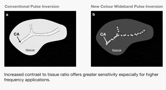
Colour Wideband Pulse Inversion (CWPI) mode offers additional contrast to tissue signal sensitivity with a choice of displays: contrast enhancement only, contrast enhancement as a colour overlay of the fundamental low MI image
MC-MTI is an accumulation mode that provides extremely detailed micro-vessel morphology visualisation, offering the potential for improved tumour characterisation. Intermittent or manual flash-replenishment sequences can be customised for each examination.
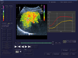
Time Intensity Curves
Time Intensity Curves (TIC) incorporating Inflow-time Mapping (ITM) can be generated from stored clips and used to display the pattern of signal enhancement with time after injection, offering improved tumour characterisation and potential for monitoring treatment response. Within multiple selectable ROIs, parameters such as time-to-peak and amplitude of enhancement, can be derived using a best-fit analysis – and all data can be exported to an Excel file for further off-line analysis.
Using the Inflow-time Mapping quantification and TIC display the earlier arrival of the contrast agent in the small liver lesion (red ROI) is displayed